

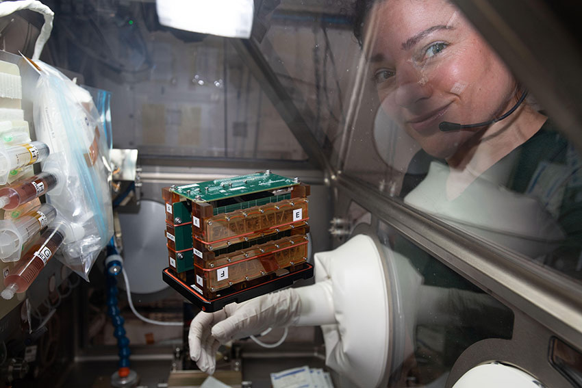
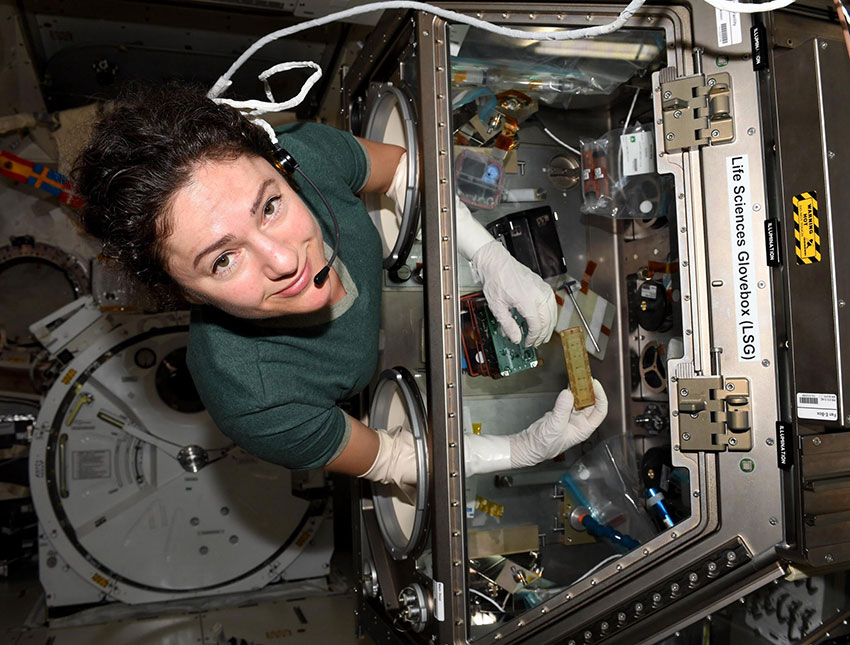
Astronaut Jessica Meir with the engineered heart tissues in their electronic habitat aboard the International Space Station (ISS). Photo courtesy of NASA
During extended space travel, astronauts can experience accelerated bone loss, changes in gene expression, and compromised immune systems and organ function. So if space programs want to make longer and further travel possible — to Mars, for example —they will need to know how to keep crews healthy.
Organ and tissue chips give researchers a unique opportunity to study the impact that space travel has on the human body. In 2016, the National Institutes of Health (NIH) partnered with NASA and the International Space Station (ISS) to launch the Tissue Chips in Space program. This spring, heart tissue from the UW joined an elite group of space travelers: chips modeling kidneys, lungs, bone and cartilage for experiments aboard the ISS.
Nathan Sniadecki
ME professor Nathan Sniadecki leads the research team behind these heart tissue chips. Over five years ago, his lab invented magnetic technology to measure the beating of these heart tissue chips. He began working with Bioengineering (BioE) faculty member Deok-Ho Kim at the UW Medicine Institute for Stem Cell & Regenerative Medicine (ISCRM) to use this technology for the ISS project. Kim has since left UW and joined the Johns Hopkins University faculty, but the work still continues in Sniadecki's lab at the UW with the help of ME doctoral student Ty Higashi and BioE postdoc Jonathan Tsui.
To determine how microgravity affects human heart muscle, the team sent their 3D beating heart tissue to the ISS via a SpaceX Dragon cargo spacecraft on March 9. The experiment ran on board the space station for a month before its successful return to Earth.
The College of Engineering’s Chelsea Yates spoke with Sniadecki — who currently serves as interim director of UW Medicine’s Heart Regeneration program — about organ and tissue chip research in space and where this work is headed next.
What exactly are organ and tissue chips?
Video courtesy of UW Medicine
Organ and tissue chips are small devices lined with living human cells. Basically they are tiny 3D cross-sections of organs and tissues. They give us a way to recreate organ-like environments to better study how organs and tissues work and how they respond to disease and treatment. In this way, they are like miniature testbeds.
As tiny models, they allow researchers to study human organ function outside of the body and at the cellular scale. We can intentionally create chips with diseases to study how organs react and also use them to test and refine drug treatments. Overall, this research promises advancements in personalized medicine: Researchers could create organ and tissue chip models unique to an individual to determine the best course of treatment for his or her specific condition.
Why send heart tissue chips to space?
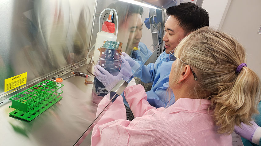
Stefanie Countryman from BioServe Space Technologies, right, helps BioE postdoc Jonathan Tsui attach tissue chambers to a circuit board to monitor the live beating of tissues while in space. Photo courtesy of Devin Mair
We know that astronauts are at risk for changes in their cardiac function and rhythm. Plus, microgravity is known to speed up aging and likely influence other cell or tissue properties. Because aging is accelerated in space, studies on the ISS allow us to assess this process over weeks instead of years. So, to learn how microgravity and other physical forces in space affect heart muscle in particular, my team packed our heart tissue chips for space.
There are Earth benefits, too. This work allows us to gather information to help prevent and treat heart muscle damage in people generally, as well as understand how aging changes heart muscle. Over the years, several space technologies — mini-cameras like those in smartphones, scratch-resistant lenses, wireless headsets and more — have found their way to life on Earth. It will be great to add tissue research like this to that list.
Tell us more about your team’s involvement in the Tissues in Space project.
Astronaut Jessica Meir with an engineered heart tissue chamber aboard the ISS. Photo courtesy of NASA
Our tissue traveled to the ISS in small, compact devices, each about the size of a smartphone. Each holder contained a row of tiny, 3D strings of beating heart tissue grown from human cells. In space and on Earth, our heart muscle tissue is supported between two flexible posts that allow it to contract freely, in contrast to the rigid confinement of a Petri dish. The posts contain tiny magnets so when the tissue contracts, the position of the magnets changes, which we can detect with a sensor. This part — embedding magnets into the post design and using magnetic detection — has been a key contribution that our team has made to heart tissue chip development.
Each of the tissue strands contains about half a million cells that act like heart tissue in the body. So it’s possible to watch these “heart strings” shorten and expand in the dish, just like little heart beats. In space that motion was detected by a sensor, and the data was sent back to our lab so we could monitor it. We also had a simultaneous twin study underway in our lab so we could compare the space tissue to tissue here on Earth.
What happens next?
There’s a lot of data, which we’ll be reviewing over the next few months so we can get a clearer picture of what’s happening to heart muscle cells and tissues in astronauts during a space mission. We plan to send our tissue to space again in the future, next time to investigate if medications and medical interventions can offset what the heart endures during extended space missions.
To learn about more research projects that explore how mechanics affect the human body and disease at the cellular level, visit Sniadecki’s Cell Biomechanics Lab.
Originally published May 11, 2020